



Engineers, Clinicians Create Training Model for Fetoscopic Repair of Spina Bifida
UC Davis Health will soon offer more treatment options for spina bifida, thanks to a team of development engineers at UC Davis Tech Foundry and medical professionals at UC Davis School of Medicine. The interdisciplinary team has created a teaching model to train fetal surgeons on fetoscopic repair of spina bifida.
Spina bifida is a congenital condition where spinal tissue fails to fuse properly in the womb, leading to lifelong health problems for approximately 1 in every 3,000 babies born in the United States. Fetoscopic repair is a minimally invasive surgical procedure to address conditions like spina bifida in a fetus. It is a type of laparoscopic surgery, where tiny incisions are made to insert very small surgical tools and a long, thin camera to help the surgical team see inside the patient’s body.
“Fetal surgery, and fetoscopy for spina bifida in particular, is one of the more technically complex procedures in fetal surgery, yet there are very few opportunities for fetal surgeons and pediatric neurosurgeons to practice it,” said Payam Saadai, a professor of surgery at UC Davis Health who had the initial idea for a training model.
One of the biggest benefits of fetoscopic repair over other surgical treatments for spina bifida is that it allows the mother to have a traditional birth in the future, due to its minimally invasive nature.
From Idea to Rough Sketch to Fully Realized Tool
Saadai passed his idea onto laboratory supervisor Christopher Pivetti and surgical research resident Elizabeth Reynolds, who both work in Professor of Biomedical Engineering and Surgery Aijun Wang’s lab at Aggie Square. Aggie Square is UC Davis’ innovation district for researchers, industry professionals and entrepreneurs to work side-by-side in Sacramento, California.
On the first floor of the district’s flagship building is Tech Foundry, a 7,500-square-foot device development facility. The space is nearly enveloped in large windows on all sides, revealing futuristic machines and engineers engrossed in work, making it hard for passersby at Aggie Square, like Pivetti and Reynolds, not to become curious about what’s going on inside.
When Pivetti and Reynolds had more time to sit and think about Saadai’s idea, they sketched out notes and diagrams on graph paper, bringing in pediatric neurosurgeons Cameron Sadegh and Marike Zwienenberg, members of the multidisciplinary UC Davis Health Center’s fetal surgery team, for their expertise in fetal closure of spina bifida as they developed the idea for the fetoscopic model. They then brought their sketch to Tech Foundry to see if the team there could bring the idea into reality.
For Valerie Quiroz, the Tech Foundry development engineer who took on the project, the sketch was all she needed. She transformed the sketch and medical jottings into a working prototype featuring 3D-printed, silicone-cast and laser-cut components.


A Teaching Model
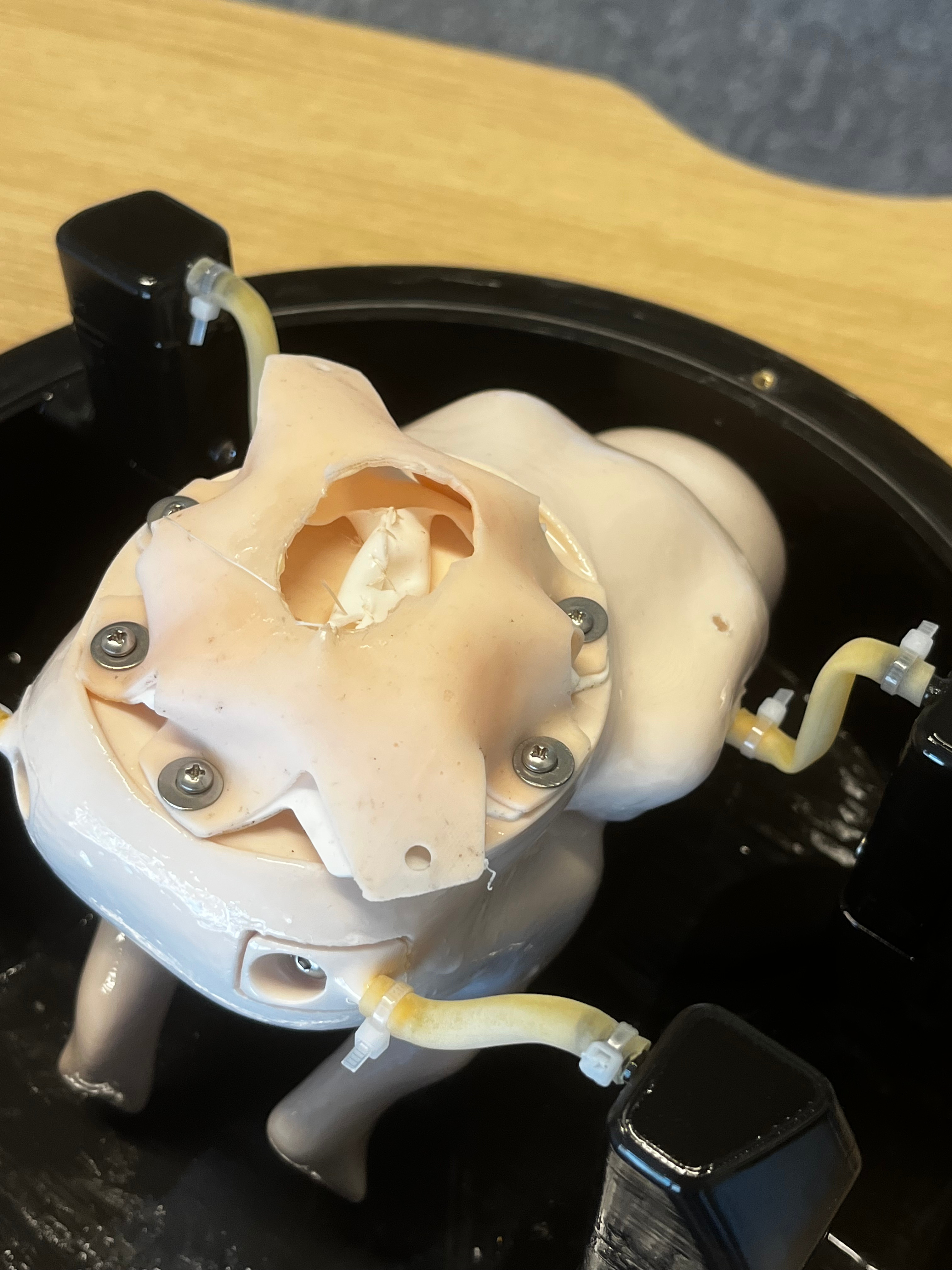
Comprising a chamber that is fillable with liquid and holds a small doll, the model simulates a fetus suspended in amniotic fluid inside the uterus. It also features a dome with small holes through which laparoscopic instruments can fit.
On the backside of the doll, or fetus model, is a raised area of skin, almost like a balloon. This is to mimic fetal myelomeningocele, the most severe form of spina bifida. It’s when the fetus’s spine and spinal canal remain open at the bottom of the spinal column, creating a bulge of skin.
“The raised area will be a consumable part that will be replaced for the next user,” Quiroz said. “It is intended to mimic multiple layers of tissue, such as skin, dura mater and placode, and allow surgeons to practice surgical techniques, including suturing.”
Due to how small the area of skin is on the fetus, coupled with how fine the laparoscopic tools are, fetoscopic repair is one of the most technical and delicate pediatric procedures. With the fetoscopic repair teaching model, fetal surgeons at UC Davis Health can now practice and perfect the technique for treating spina bifida before ever entering the operating room.
“The fetal operation requires technical precision and is only available at very few centers in the world,” Saadai said, “so the fetal model directly supports this work by creating a safe, reproducible environment where our team can train and refine these techniques, dramatically shortening the learning curve for such a complex operation.”
Advancing the CuRe
Thanks to the teaching model, fetoscopic surgery will be an option for spina bifida treatment alongside the CuRe trial at UC Davis Health. The CuRe trial, or Cellular Therapy for In Utero Repair of Myelomeningocele, is a groundbreaking treatment using a stem cell patch to repair spina bifida tissue.

The trial is led by Professor of Surgery Diana Farmer and Professor of Biomedical Engineering and Surgery Aijun Wang, with Pivetti, Reynolds and Saadai as members of the clinical research team, among many others. The trial is a world first and was recently found to be safe in a paper published in The Lancet, but it requires invasive surgery.
The training model will be used to test the next stage of the CuRe Trial, which is to transform it into a minimally invasive fetoscopic surgery, where a surgeon would administer the small stem cell patch with laparoscopic tools before closing the fetus’s skin.
“By developing not just fetoscopic techniques but also the training infrastructure, which we have never had at UC Davis before, we position our health center as one that is advancing the next generation of both fetal therapy and surgical innovation,” Saadai said.