UCD BME Professor Simon Cherry's Lab Part of Team That Created Cross-Sectional Tissue Scans Without Tomographic Reconstruction
Superfast photon detectors image at the speed of light
Quick Summary
- This direct imaging technique promises to make PET/CT scans safer, easier, quicker and more affordable
Current medical imaging techniques such as CT and PET scans can be time-consuming, expensive and involve cumbersome equipment. One reason why they are so laborious is the need for tomographic reconstruction, where a mathematical algorithm is used to create a cross-sectional image from the raw data. Now, researchers in the U.S. and Japan have produced the first cross-sectional image of tissue without the extra step of tomographic reconstruction. The work was published Oct. 14 in the British scientific journal Nature Photonics.
The advance was made possible by development of new, ultrafast photon detectors, according to Simon Cherry, professor of biomedical engineering (BME) and radiology at UC Davis and senior author on the paper.
“We’re literally imaging at the speed of light, which is something of a holy grail in our field,” Cherry said.
Experimental work was led by Sun Il Kwon, project scientist in UC Davis’ BME department and Ryosuke Ota at Hamamatsu Photonics, Japan. Other collaborators included research groups led by Professor Yoichi Tamagawa at the University of Fukui, and by Professor Tomoyuki Hasegawa at Kitasato University.
In positron emission tomography (PET) scans, molecules tagged with trace amounts of a radioactive isotope are injected and taken up by the body. The isotope, often fluorine-18, is unstable and emits positrons as it decays.
Whenever one of these positrons encounters an electron within any of the atoms in the body, the positron and electron ‘annihilate’ each other and simultaneously give off two ‘annihilation’ photons. Theoretically, researchers could reconstruct the origin and trajectory of these annihilation photons to identify the location of atoms in the body and thus construct an image of the tagged molecules in the tissue. But until now, researchers were unable to do that without the extra step of tomographic reconstruction because detectors were too slow to precisely determine the arrival times of the two photons.
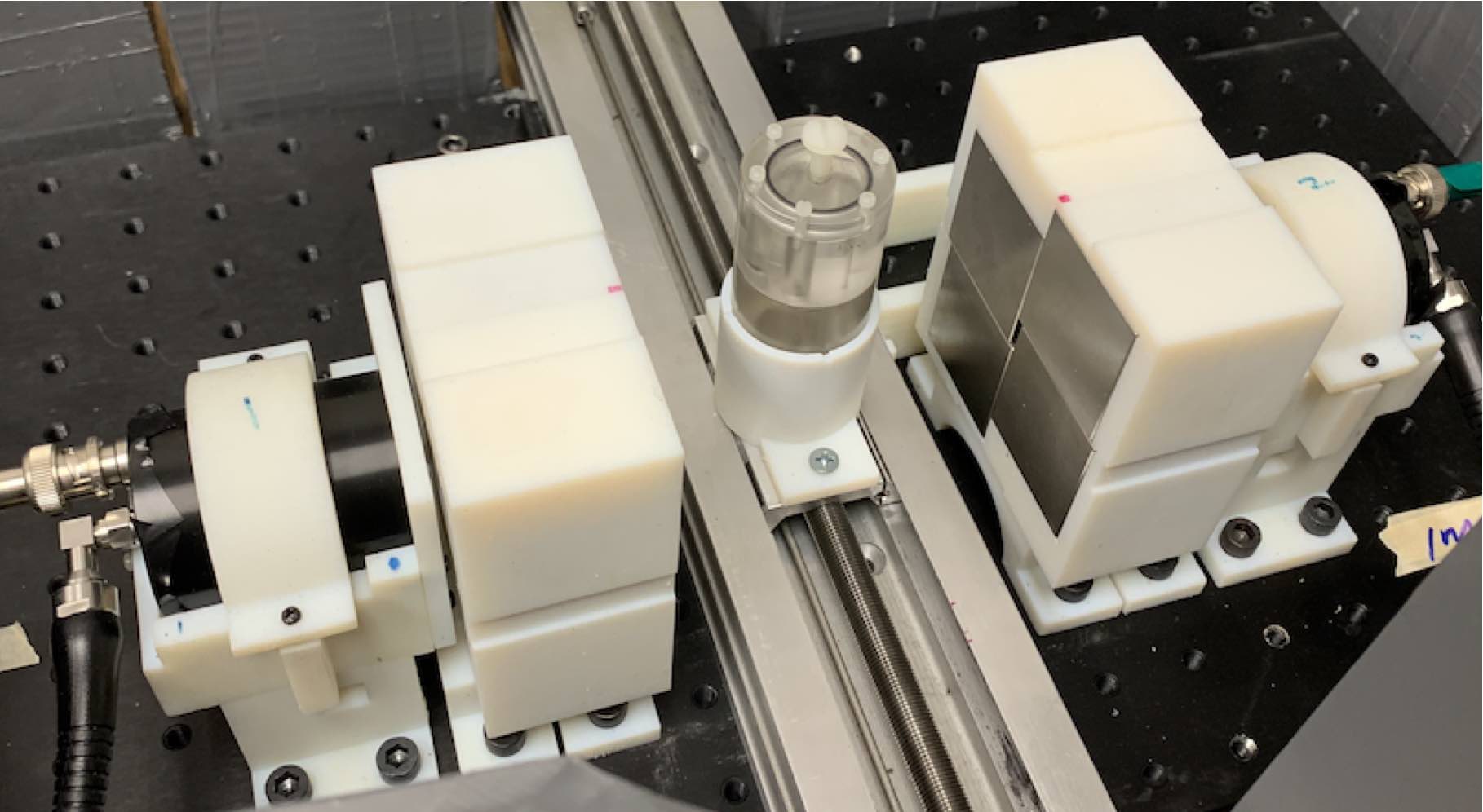
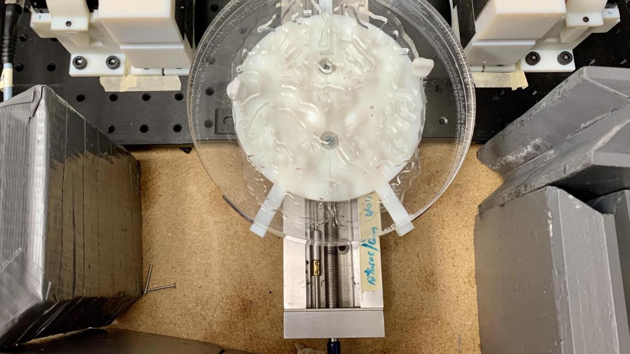
Cherry and his fellow researchers figured out how to detect super-fast Cherenkov photons, emitted when annihilation photons interact in suitable materials, with an average timing precision of 32 picoseconds. They can determine where the annihilation photons arose with a spatial precision of 4.8 millimeters. This level of speed and accuracy enabled the research team to produce cross-sectional images of a positron-emitting radionuclide directly from observing the coincident annihilation photons, without having to use an algorithm for tomographic reconstruction.
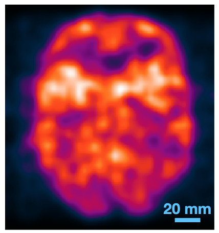
In their paper in Nature Photonics, the researchers describe various tests they conducted with their new technique, including with a test object that mimics the human brain. They feel confident that this procedure is ultimately scalable to the level needed for clinical diagnostics and has the potential to create higher quality images using a lower and safer radiation dose. Images can also be created much more quickly with this method, potentially even in real time during the PET scan as no after-the-fact reconstruction is needed.
PET scans are currently quite expensive and are also technically limited by the need for the reconstruction step. This new breakthrough involves a compact equipment setup and offers the promise of cheaper, easier, and more accurate scans of the human body using radiotracers.