Imaging the Living Brain Unlike Ever Before
New technology improves on two-photon microscopy, making it easier to study neuroplasticity
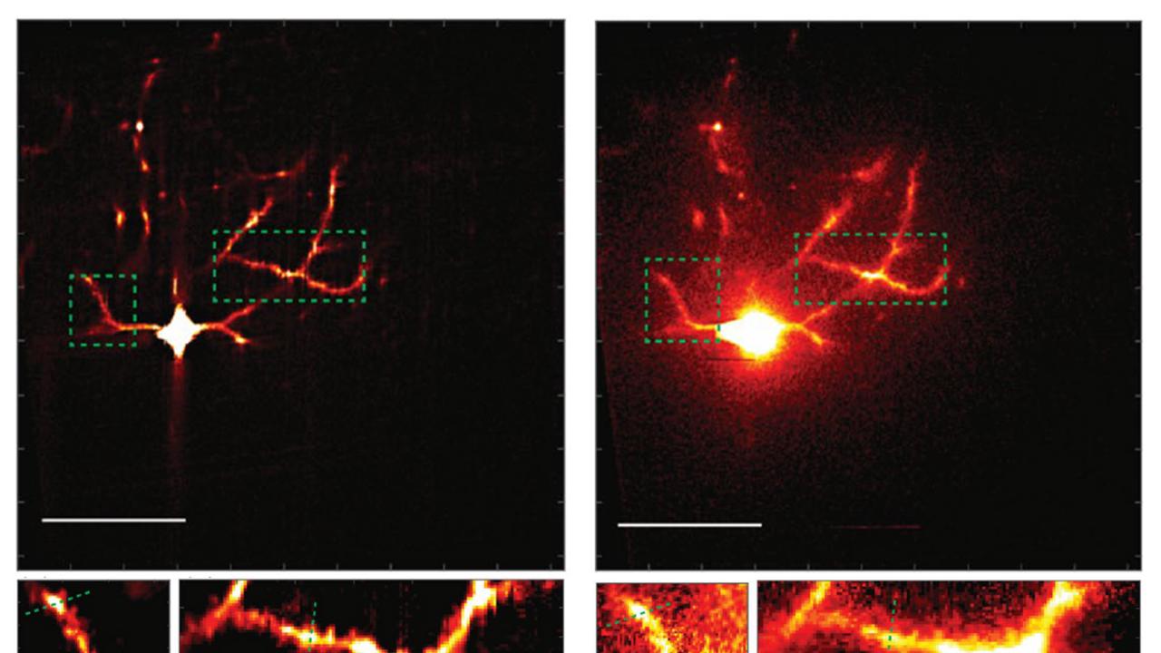
Assistant Professor of Biomedical Engineering Yi Xue is the lead author of a paper, published May 13 in Nature Scientific Reports and in collaboration with researchers from MIT, that describes a new microscopy system that significantly improves upon the traditional two-photon method for in vivo imaging of the brain.
Called mosTF, short for multiline orthogonal scanning temporal focusing, the system is eight times faster and four times clearer than two-photon imaging. The increased speed and clarity enable detailed tracking of living brain tissue unlike ever before, promising to provide new insights into neuroplasticity, or the brain’s ability to adapt, learn and rewire itself.
The technology works by scanning brain tissue with lines of light in perpendicular directions. This method is different from other imaging techniques that go point by point.
“Our excitation light is a line rather than a point — more like a light tube than a light bulb,” Xue said.