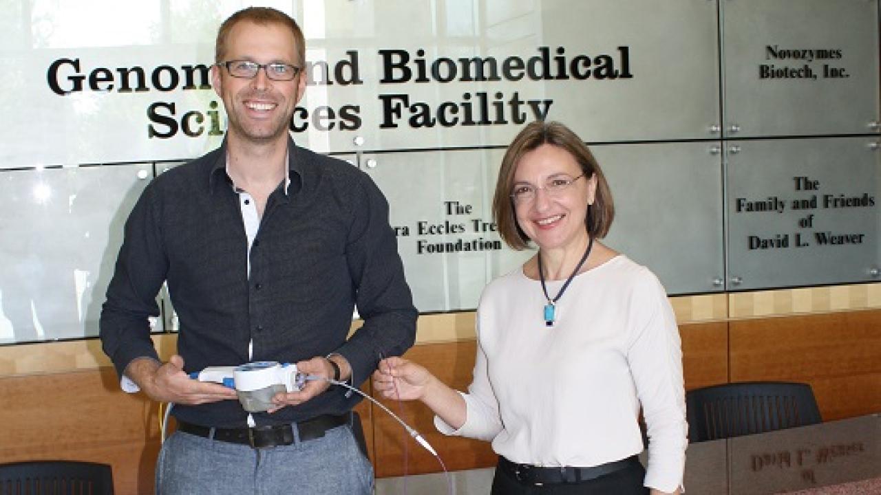
New imaging catheter is first of its kind in war against heart disease
The catheter used in the study is flexible enough to access coronary arteries in a live human following standard procedures, and does not require injected fluorescent tracers.
To win the battle against heart disease, cardiologists need better ways to identify the composition of plaque most likely to rupture and cause a heart attack. Angiography allows them to examine blood vessels for constricted regions by injecting them with a contrast agent before x-raying them. But because plaque does not always result in constricted vessels, angiography can miss dangerous buildups of plaque. Intravascular ultrasound (IVUS) can penetrate the buildup to identify depth, but lacks the ability to identify some of the finer details that convey information about risk of plaque rupture. With a better tool that combines the strengths of depth and biochemical imaging, cardiologists will have more success to prevent heart attacks.

For the first time, Laura Marcu’s biomedical engineering lab at UC Davis has combined intravascular ultrasound (IVUS) with fluorescence lifetime imaging (FLIm) in a single catheter probe capable to image the tiny arteries of a living heart. The new catheter can simultaneously retrieve structural and biochemical information about arterial plaque that could more reliably predict heart attacks.
In a paper published August 21, 2017 in Scientific Reports, Marcu’s group describes construction of the first device that integrates a fiber optic and an ultrasound probe within an intravascular catheter to simultaneously analyze the chemical composition and the morphological features of the narrow coronary arterial vessels in a beating heart.
Short laser pulses are sent through the fiber optic to excite specific molecules in tissue, which releases a tiny amount of light in return. The intensity and duration of the emitted light depends on the biochemical makeup of the tissue such as the amount collagen, elastin, or lipids. When combined, the FLIm-IVUS imaging catheter can provide a comprehensive insight into how atherosclerotic plaque forms, diagnostics, and response to therapy.
The new catheter was tested in living swine hearts and samples of human coronary arteries.
The catheter used in the study is flexible enough to access coronary arteries in a living human following standard procedures. The team also overcame the interference of heart motion with the probe’s ability to rapidly collect data. They used a transparent solution to displace blood to get a clean view of the artery walls. Importantly, this imaging catheter system does not require injected fluorescent tracers or any special modification of the catheterization procedures.
The researchers believe that this new intravascular diagnostic technology may benefit patients and researchers alike. In vivo evaluation of plaque with this FLIm-IVUS technique not only can improve understanding of mechanisms behind plaque rupture – an event with fatal consequences- but also the diagnosis and treatment of patients with heart disease. Marcu’s group is currently working to obtain FDA approval to test this new intravascular technology on human patients.
In vivo label-free structural and biochemical imaging of coronary arteries using an integrated ultrasound and multispectral fluorescence lifetime catheter system. Julien Bec, Jennifer E. Phipps, Dimitris Gorpas, Dinglong Ma, Hussain Fatakdawala, Kenneth B. Margulies, Jeffrey A. Southard & Laura Marcu. Scientific Reports 7, Article number: 8960 (2017). doi:10.1038/s41598-017-08056-0
The videos below show how the catheter visualizes the arteries.
