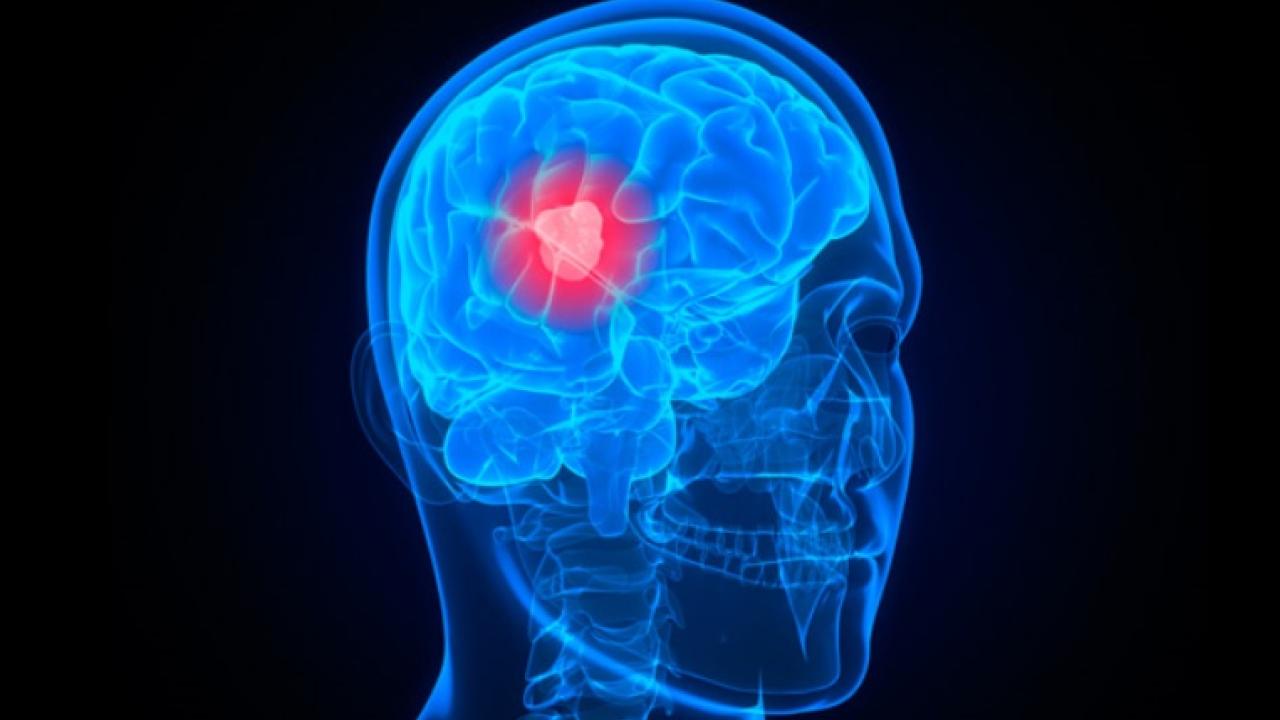
Brain Innovation Group Funded for Brain Tumor Tool
The UC Davis Comprehensive Cancer Center’s Brain Innovation Group has received a grant from the National Cancer Institute (NCI) to improve brain cancer surgery and treatment using UC Davis-developed biophotonic technology.
The $400,000 grant is the first for the cancer center’s eight cancer research innovation groups, which link scientists, oncologists, surgeons, engineers and other experts in discussions about patient care needs and potential innovations.
“The groups were started to fulfill a big part of our mission as a comprehensive cancer center by enhancing clinical and translational cancer research,” said cancer center director Ralph de Vere White. “This grant is a clear example of the success of this endeavor.”
UC Davis researchers will use the funding to adapt state-of-the-art optical biopsy technology, the Multispectral Scanning-Time Resolved Fluorescence Spectroscopy, to help neurosurgeons distinguish between radiation necrosis and cancer recurrence during brain cancer surgery. The technology was developed by Laura Marcu, professor of biomedical engineering and neurological surgery and principal investigator on the project.
The collaborative Brain Innovation Group includes specialists from adult and pediatric oncology, neurology, neurosurgery, neuroradiology, radiation oncology, biomedical engineering and biophotonics, hematology and biochemistry. They meet once a month in an open forum to present their projects and look for ways to combine and translate their work into high-impact clinical trials.
“This NCI grant demonstrates the benefit of having experts with different backgrounds work together to find new ways to better diagnose and treat cancer,” said Marcu, adding that the idea to apply the novel photonic technology in distinguishing between brain tumor recurrence and radiation necrosis was sparked during an innovation meeting.
Brain tumor expert Fredric Gorin said a tool to help distinguish different types of brain tissue is needed in the field of brain tumor diagnosis and treatment.
“The brain is very sensitive to radiation damage,” said Gorin, chair of the UC Davis Department of Neurology and project adviser. “When radiation necrosis happens in the area of brain cancer, it is difficult to distinguish which tissue represents recurrent brain cancer and which is tissue damaged by radiation. The MRI often can’t distinguish between the two.”
Because many brain and spinal cord tumors recur, it’s common for brain cancer patients to have both recurrent and necrotic tissue, Gorin explained. Having a way of distinguishing between the two can have a significant impact on treatment options and patient survival.
“It makes all the difference in the world to know,” said Ruben Fragoso, a UC Davis radiation oncologist, who will generate necrosis specimens from rat models for the study. “If the tissue is necrotic from radiation, the last thing you want to do is give more radiation.”
Marcu’s diagnostic optical device uses laser light to excite molecules within tissues. She hopes it will reveal with pinpoint-accuracy the biochemical status of the tissue in real-time during surgery without the need to inject any contrast agents. If successful, this approach could dramatically improve the precision of surgical techniques, serve as an invaluable diagnostic tool and play a big role in optimizing radiation therapy.
“If this tool can help surgeons better define where there is more tumor, we can potentially fine-tune radiation treatment plans by knowing which areas need greater margin coverage or possibly more radiation,” Fragoso added.
The study is underway in rat models. The goal is to launch human clinical trials next year, under the guidance of James Boggan, chair of the UC Davis Department of Neurological Surgery.
The Brain Innovation Group co-chairs are medical oncologist Robert O’Donnell and Paul Knoepfler from the UC Davis Department of Cell Biology and Human Anatomy.
Story source: http://www.ucdmc.ucdavis.edu/publish/news/newsroom/9287
