Using Imaging Tools to Treat Pancreatic Cancer
Biomedical engineering professor Julie Sutcliffe and her team are using their expertise in cancer imaging and diagnosis to develop a new, effective treatment for pancreatic cancer. After her team created a therapeutic based on a radioactive imaging tool they previously developed, the team received $4 million from the Lustgarten Foundation and Stand Up to Cancer to bring their drug to clinical trials to help patients suffering from the disease.
Pancreatic cancer is a particularly deadly and difficult cancer to treat. It’s often asymptomatic until very late stages, and the pancreas itself is very dense, making it impossible to deliver drugs directly to the source. The only true treatment is surgically removing part or all of the pancreas, but the surgery can’t be done if the cancer has metastasized. The goal, then, is to find and eradicate the metastasized tumors outside the pancreas to make the surgery an option for more patients.
“Surgery is really going to be the only cure, so if you can’t have surgery because you have metastatic disease, then we’re going to use this therapy to treat that metastatic disease first so they can operate,” she said.
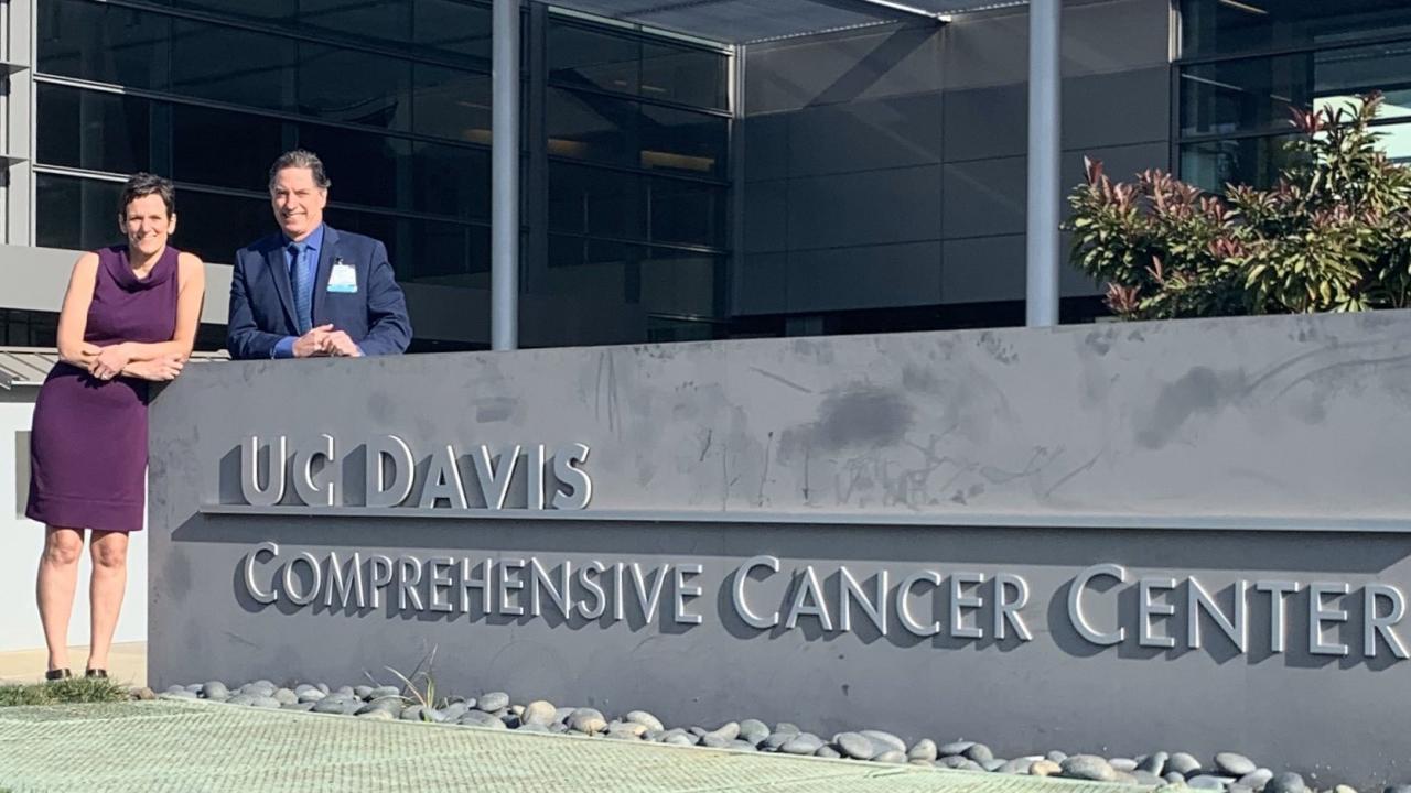
Sutcliffe’s work in this area began 15 years ago, when she herself was being treated for breast cancer. She and her surgeon, Dr. Richard Bold, began discussing their work and realized they needed each other’s expertise in imaging and pancreatic cancer, respectively. The two have been frequent collaborators since and have made significant progress in accurately imaging and diagnosing metastasized pancreatic cancer.
To do this, they developed radioactive molecules in their lab that bind to receptors that only exist on metastasized pancreatic cancer cells. These molecules allow them to accurately image the disease using a PET scanner—something some experts didn’t think was possible.
In 2018 when the Lustgarten Foundation and Stand Up to Cancer called for proposals that fast-tracked therapeutics for pancreatic cancer, Sutcliffe and her team took the challenge in stride to turn their diagnostic tool to treatment. The tool still attaches to the cell, but it now kills it instead of simply lighting it up for the PET scanner.
“When they said to us ‘you have a year to develop a therapeutic,’ we said, ‘ok, game on,’” said Sutcliffe. “If I change the one radioactive isotope on my diagnostic, I now have a radionuclide therapy [tool] that I can use to treat.”
The team, which includes her surgeon collaborator and several of their colleagues at the UC Davis School of Medicine, received $1 million in 2018 to develop the tool and conduct initial pre-clinical tests. After finding success, they were rewarded for their efforts with the three-year, $4 million extension, which they will use to conduct clinical trials in human patients.
“We’ve got all the preclinical data, we have the toxicology package and we make the drug in my lab,” she said. “So now not only does UC Davis have this first-in-human diagnostic, but they will also have a first-in-human therapy that was homegrown out of our lab.”
Sutcliffe stresses the importance of her team, both in her lab and at the UC Davis School of Medicine. The team brings together chemists and biologists in her lab with surgeons, pathologists, radiation and medical oncologists and nuclear medicine physicians who all contribute expertise and experience to all aspects of developing this new treatment.
“It’s team science,” she said. “Imagine the person power it took to get from an idea—we didn’t even have the drug—through to preclinical testing to enough data to get an IND application in a year. My team is so diverse and I need all of them.”
